Who Knew Glowing DNA Could Look So Awesome?
Feb 06, 2014 18:59
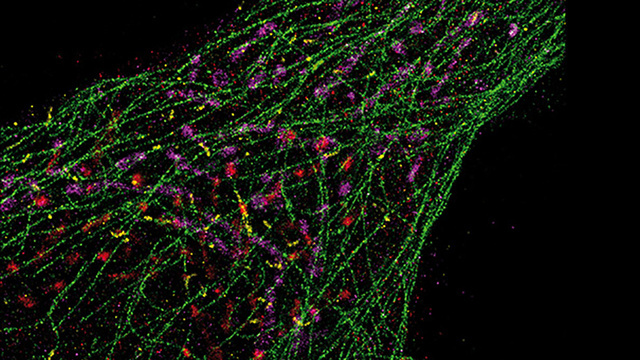
Nope, this is not some trippy light show you're looking at. It's actually a single human cell that's been lit up using a new method called Exchange-PAINT. Created by Harvard researchers, this fascinating technique uses DNA's specific binding characteristics to create ultra-sharp images of structures that are too small to be seen with traditional light microscopes.
The process involves coating the structures inside the cell with unique DNA tags. Free-floating DNA carrying these fluorescent molecules are then added, and each color is coordinated to a different type of DNA tag. When the fluorescent DNA labels attached themselves, the result was an image with resolution less than 10 nanometers—one-twentieth the diffraction limit of a light microscope.
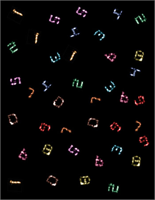
The researchers have managed to create 10 unique pieces of tiny folded DNA shaped like the numbers 0 through 9. The next phase is to refine the process in order to create ultra-precise images that go on inside living cells which are currently too small to directly observe.


